© estudio pobrelavaca 2006


  1 

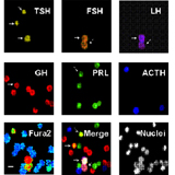
BIOLUMINISCENCE IMAGING OF SUBCELLULAR CALCIUM USING TARGETED AEQUORINS.

Subcellular calcium analysis has been enabled by the introduction of targeted, protein-based Ca2+ probes as for example aequorin. However, studies at the single cell level have been hampered by the low light level of aequorin. Photon counting imaging of cells expressing high levels of targeted aequorins allows now the monitoring of Ca2+ in mitochondria, nucleus, or whatever organelle at the single cell level.


  2 

REPORTER GENE EXPRESSION IN SINGLE, LIVING CELLS BY PHOTON COUNTING IMAGING OF LUCIFERASE REPORTER GENE.

Dynamics of gene expression in individual cells can be performed by imaging bioluminescence from individual cells expressing the luciferase reporter gene under control of an entire gene promoter or specific response elements for transcription factors and nuclear receptors.


  3 

CALCIUM IMAGING BY DIGITAL FLUORESCENCE MICROSCOPY.

Analysis of cytosolic calcium changes in individual cells is carried out by conventional fluorescence microscopy of cells loaded with ratiometric dyes fura-2 or fura4F.


  4 

MITOCHONDRIAL POTENTIAL IMAGING BY FLUORESCENCE AND CONFOCAL MICROSCOPY.

Mitochondrial potential can be monitored in intact cells by fluorescence microscopy of cells loaded with fluorescent probes that tend to accumulate in mitochondria as TMRE and JC1. Confocal microscopy allows to disect between mitochondria and the surrounding cytosol.


  5 

MULTIPLE SEQUENTIAL IMMUNOCYTOCHEMISTRY.

The anterior pituitary is made of 5 different cell types expressing 6 different pituitary hormones. Some cells may store multiple hormones. We have established a new method for simultaneous 6 fold immunocytochemistry to type anterior pituitary cells for all the hormones they stored. The method can be used for any other antigen and can be combined with calcium imaging to establish functional responses to agonists in the same single cells.