| MULTIPLE SEQUENTIAL IMMUNOCYTOCHEMISTRY. |
|
cells kept in the microscope stage were fixed with 4% paraformaldehyde in phosphate buffered saline (PBS), permeabilized with 0.3% triton X-100 and washed with PBS. Then 10% goat serum in PBS was added. After 5 min., cells were incubated with antibodies against three human AP hormones (TSH, FSH, LH) labeled with Oregon Green 488, Cascade Yellow and Alexa 350, respectively. After washing, specific fluorescence images corresponding to each fluorophore were captured to reveal stained cells with the following fluorescence settings: Oregon Green (FSH): excitation, 490 nm; emission, >510 nm); Cascade Yellow (TSH) excitation, 380 nm, emission, >510 nm; Alexa 350 (LH), excitation, 340 nm; emission, >450 nm. This step enables typing cells storing either TSH, LH, FSH as well as cells co-storing combinations of these AP hormones. Once the first series of images were captured and stored, cells were washed and incubated again with antibodies against GH, PRL and ACTH labeled with Oregon Green 488 (PRL), Cascade Yellow (GH) and Alexa 350 (ACTH), respectively, and the incubation was continued for 30 min. Then cells were washed and three new fluorescence images were taken with the same fluorescence settings described above. This new series of images revealed cells stained by the first antibody plus those newly stained by the second one. Cells stained by the second series of antibodies were revealed by subtracting the first series from the second one. Figure shows the merger of the staining with the different antibodies in a representative experiment. Finally, nuclei were stained with Hoechst 33258 (0.5 µg/ml, 10 min) and another fluorescence image was acquired (excitation, 340 nm; emission, >420 nm). The nuclear images allows to distinguishing individual cells that were physically close.
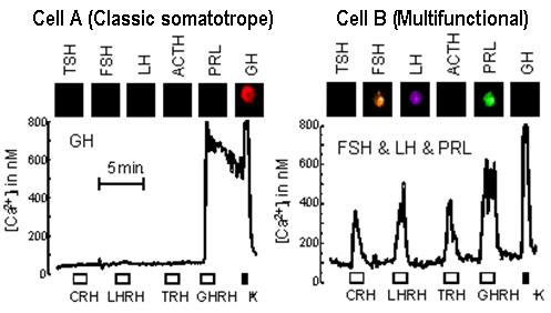
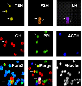 Anterior pituitary cells were loaded with fura2 and subjected to calcium imaging to reveal functional responses to multiple hypothalamic releasing factors. Then, cells were fixed and six-fold immunocytochemistry against the 6 anterior pituitary hormones was carried out in the same cells. Finally, nuclei were also stained. Thus, expression of up to four hypothalamic releasing hormone receptors and six anterior pituitary hormones was assessed simultaneously in single cells. Arrows point to cells storing more than one hormone. Examples of such phenotypes are shown. Cell A stored only GH and responded only to GHRH (a classic somatotrope). Cell B stored multiple hormones (FSH,LH and PRL) and expressed all the four classic hypothalamic releasing hormone receptors (a multifunctional phenotype). Anterior pituitary cells were loaded with fura2 and subjected to calcium imaging to reveal functional responses to multiple hypothalamic releasing factors. Then, cells were fixed and six-fold immunocytochemistry against the 6 anterior pituitary hormones was carried out in the same cells. Finally, nuclei were also stained. Thus, expression of up to four hypothalamic releasing hormone receptors and six anterior pituitary hormones was assessed simultaneously in single cells. Arrows point to cells storing more than one hormone. Examples of such phenotypes are shown. Cell A stored only GH and responded only to GHRH (a classic somatotrope). Cell B stored multiple hormones (FSH,LH and PRL) and expressed all the four classic hypothalamic releasing hormone receptors (a multifunctional phenotype).
|
| |
|
For further details see:
|
Multifunctional cells in human pituitary adenomas: implications for paradoxical secretion and tumorigenesis. |
| |
| Senovilla L, Núñez L, de Campos JM, de Luis DA, Romero E, Sánchez A, García-Sancho J, Villalobos C (2004). J Clin Endocrinol Metab 89, 4545-4552. |
| |
|
|
|
Multifunctional cells of mouse anterior pituitary reveal a striking sexual dimorphism. |
| |
| Núñez L, Villalobos C, Senovilla L, García-Sancho J (2003). J Physiol (London) 549, 835-843. |
| |
|
|
Back>>
|